University of Delaware
![]()
College undergraduates were identified as alexithymic or control, based on their scores on the Toronto Alexithymia Scale (Taylor, Ryan, & Bagby, 1985). All subjects were presented standardized emotion-eliciting color slides for six seconds while facial muscle, heart rate, and skin conductance activity were recorded. Stimuli were presented a second time while subjects were asked to provide emotion self-reports using a paper and pencil version of the Self-Assessment Manikin (SAM; Lang, 1980) and to generate a list of words describing their emotional reaction to each slide. Consistent with the definition of alexithymia as a syndrome characterized, in part, by a deficit in the identification of emotion states, high TAS subjects supplied fewer emotion-related words than did controls to describe their response to the slides. Alexithymics also indicated less variation along the arousal dimension of the SAM, produced fewer specific skin conductance responses and showed less heart rate deceleration to the slides, regardless of category. No valence-related differences between alexithymic and control subjects were noted.
![]()
Sifneos (1972) coined the term alexithymia to designate a group of cognitive and affective characteristics typical of many patients with psychosomatic illnesses. It is thought to be a personality trait that is characterized by a decreased ability to communicate feelings, a decreased ability to identify feelings, a cognitive tendency toward detail and external operations or events, and a paucity of imaginative thought, dream recall, or fantasy (Taylor, 1994).
is directly discharged through outward expression or is indirectly expressed through internal pathways. According to discharge theory, outward expression leads to an attenuation of physiological reactivity. Conversely, when emotions are not expressed (discharged) outwardly, they will be expressed in terms of immoderate sympathetic activation. It follows from this theory that alexithymics would have a higher probability for excessive sympathetic activity during emotion, given that the ability to communicate emotions includes an outward expression of affect to others and that alexithymics are characterized by a deficit in emotional communication. Because a stress response entails sympathetic arousal, an intensification of this response due to emotional energy which is not outwardly expressed might hypothetically lead to excessive or prolonged activity of organs innervated by the sympathetic nervous system and, in turn, lead to tissue degeneration and consequent somatic illness.
Martin and Pihl (1985), in their Astress-alexithymia hypothesis@, also advocated a causal relationship between alexithymia and psychosomatic illnesses. According to this hypothesis, alexithymics Alack the affective awareness which would permit identification of a particular situation as stressful,@ (p.170) and therefore experience stressful events longer and more frequently than non-alexithymics. Although the mechanism discussed by Martin and Pihl (i.e., lack of emotion modification) differs from that in the discharge theory (i.e., conservation of energy), the effect of stress on alexithymics is similar in the two views. Both suggest increased or excessive sympathetic activity.
The evidence from laboratory studies that have examined alexithymics' responses to stress does not, however, provide unequivocal support for the prediction of increased sympathetic activity. Although some authors have concluded that alexithymics do experience elevated autonomic activity in response to stress-inducing experimental paradigms (Martin & Pihl, 1986; Martin, Pihl, Young, Ervin, & Tourjman, 1986; Rabavalis, 1987), others have found evidence which contradicts this notion and some have even concluded that alexithymics exhibit less autonomic reactivity than controls under conditions of stress (Hyer, Woods, Summers & Boudewyns, 1990; Newton & Contrada, 1994; Papciak, Feuerstein, & Spiegel, 1985). The inconsistency of these findings may be due in part to the use of various, poorly standardized measures of alexithymia. Regardless, given that the causal direction of the alexithymia-psychosomatic relationship has yet to be proven and that the defining characteristics of alexithymia do not involve stress responses but rather an affective style, it seems logical at this point to investigate alexithymics= responses to a set of stimuli that vary in affective content rather than to stimuli designed to elicit only negative affect. Such a line of investigation holds the promise of providing a broader understanding of alexithymics= affective response profile which might then, in turn, help direct future research.
Although several studies have examined alexithymics' responses to both positive and negative stimuli (e.g., Lane et al., 1996; Mayor, DiPaolo & Salovey, 1990; Mann, Wise, Trinidad, & Kohanski, 1994; McDonald & Prkachin, 1990), only two have included measures of physiological activity (Nemiah, Sifneos & Apfel-Savitz, 1977; Wehmer, Brejnak, Lumley, & Stettner, 1995). Both of these studies have areas which the present study was designed to improve upon. The Nemiah et al. experiment employed a fairly idiosyncratic methodology, identified subjects with a fairly unreliable assessment instrument, and lacked sufficient verification of mood manipulation (as discussed by Martin & Pihl, 1986). The methodology of Wehmer et al. was more standardized (cf. Lang, Bradley & Cuthbert, 1997) and included the use of the TAS for subject identification, but it was somewhat limited from a psychophysiological perspective. For example, although they collected pleasantness (valence) ratings of stimuli, along with measures of heart rate and skin conductance, they did not collect ratings of emotional arousal: a variable which has been shown to vary linearly with skin conductance (Greenwald, Cook & Lang, 1989) and which is considered to be one of the main dimensions of emotions (Abelson & Sermat, 1962; Cliff & Young, 1968; Green & Cliff, 1975; Russell, 1980; Russell & Mehrabian, 1977; Schlosberg, 1952). Furthermore, they did not collect facial electromyographic data which is generally a more reliable index of emotional valence than heart rate (Greenwald et al., 1989).
A more direct application of the methodology developed by Lang and his associates would seem to be well suited for an examination of alexithymics= physiological and self-report characteristics in response to a broad spectrum of emotion-eliciting stimuli. This well-validated methodology assesses affective behavior across multiple response systems (behavioral, verbal, and physiological) and conceptualizes emotion as a behavioral complex located in a three dimensional affective space (Lang, 1985; Lang, Bradley & Cuthbert, 1997). The three dimensions of this affective space are arousal (excited to calm), valence (pleasant to unpleasant), and dominance (controlled to in control). Studies examining facial expressiveness (Schlosberg, 1952), affective language (Russell & Mehrabian, 1977; Smith & Ellsworth, 1985), and judgements of text (Russell, 1980) have demonstrated that the majority of variance is accounted for by the dimensions of valence and arousal.
To develop reliable methods for examining emotional reactions from this dimensional perspective, Lang, Ohman, and Vaitl (1988) developed a set of slides (the International Affective Picture System; IAPS) which elicit a wide variety of affective reactions. In addition to the reliability of the self-reports elicited by the IAPS (as measured by the Self-Assessment Manikin; SAM; Lang, 1980), research has uncovered reliable relationships between a number of physiological responses and the dimensional self-reports. Greenwald et al. (1989) and Lang, Greenwald, Bradley, and Hamm (1993) found that integrated corrugator electromyogram (EMG) was inversely related to slide valence, while the relationship between valence and zygomatic EMG was direct. In addition, while skin conductance response (SCR) parameters were found to reliably index arousal self-report, relative heart rate (HR) acceleration was related to slide valence, though less strongly than the facial EMG measures. The consistency of these relationships between the physiological measures and the dimensional self-report measures has been supported by many subsequent studies (e.g., Bradley, Greenwald, Petry, & Lang, 1992; Bradley, Lang, & Cuthbert, 1993; Greenwald et al., 1989; Lang et al., 1993; Simons, Fitzgibbons, & Fiorito, 1993).
Alexithymics' self-reports to the IAPS and their EMG responses to stimuli targeting a variety of affective experience are areas which are at present uninvestigated1. Most of the evidence concerning alexithymics= responses to affect-laden stimuli concerns judgments of content rather than self-reports of experience. Some of these studies show that alexithymics judge emotional stimuli as accurately as controls, while most others report either biases and deficits related to the evaluation of negative emotions or overall lower accuracy in identifying affective stimuli (e.g., Berenbaum & Prince, 1994; Jessimer & Markham, 1997; Parker, Taylor & Bagby, 1993). The literature suggests that alexithymics may also show a deficit in spontaneous facial expressions of negative affect (McDonald & Prkachin, 1990), though their posed facial expressions appear normal. Only two studies have entailed an examination of alexithymics' self-report of emotion experience to a wide variety of affective stimuli (Lane et al., 1996; Wehmer et al., 1995). These examinations are notable in the literature because impoverished affective self-description is a definitive criterion for alexithymia and stimulus judgment deficits are not; therefore, assessment of self-descriptions is, by definition, more pertinent to assessing alexithymia than is assessment of stimulus judgments. Unfortunately the evidence is not decisive. Lane at al. (1996) found that alexithymics differed from controls in their affective self-descriptions; Wehmer et al. (1995) found no differences in positivity ratings in response to slides but did find that lower scores on two TAS factors correlated with a lower percentage of emotional words given in response to emotionally evocative scenes.
The present experiment included an examination of heart rate, skin conductance, facial muscle, and emotion self-report responses to a set of color-slide stimuli chosen to elicit affective responses. Because Wehmer et al. (1995) undertook a similar investigation of alexithymia, their results provide one of the only sets of data from which the present experiment=s results can be predicted. In Wehmer et al.=s study, alexithymics did not differ from controls in terms of their self-reports of pleasantness (i.e., valence) nor in terms of HR change in response to the affective slides across pleasantness ratings. They also found that alexithymics had a higher baseline HR than controls and that alexithymia was associated with less skin conductance responding during slide presentation.
Based on the pleasantness ratings and phasic heart-rate data reported by Wehmer et al. (1995), it could be argued that alexithymics process the valence aspects of emotion normally. Their skin-conductance data, however, suggest that the alexithymic affective deficit may be related to processing of the arousal dimension. Because Wehmer et al. did not include an arousal self-report scale, it is possible that a more targeted self-report measure might help to flesh out an arousal deficit. Such a deficit might underlie the well documented deficit evinced by alexithymics in the expression and identification of emotions.
Although Wehmer et al. (1995) did not measure facial EMG, it was included in the present study because it has a particularly robust relationship with emotion valence, and the absence of group differences in facial EMG would give added strength to the argument that processing of emotion valence is undisturbed in alexithymia.
Last, the current experiment included a more open-ended written response which required subjects to produce a list of words describing how each slide made them feel. This measure was included in order to assess one of the hallmark features of alexithymia -- i.e., a deficit in one=s ability to describe one=s emotions. On this measure, alexithymics were expected to list fewer emotional words that describe their feelings than controls, and it was expected that this might be particularly true for words used to describe high arousal emotion. This latter hypothesis was based on the supposition that Wehmer et al.'s (1995) data indicate decreased responsiveness to the arousal dimension of affect and that this might be reflected in the words alexithymics used to describe their experience.
![]()
Subjects
Subjects were selected from General Psychology classes at the University of Delaware based on scores obtained on the 26-item Toronto Alexithymia Scale (TAS; Taylor et al., 1985). The TAS is a self-report questionnaire that measures the ability to describe and identify feelings, the ability to distinguish between feelings and bodily sensations, the tendency to daydream, and the tendency to exhibit externally oriented thinking . Subjects respond to TAS items (e.g., "I have feelings that I can't quite identify") on a 5-point scale which ranges from "Strongly Disgree" to "Strongly Agree." The TAS exhibits test-retest stability (one week r = 0.82; five week r = 0.75; Taylor et al., 1985) and construct and criterion-related validity (Bagby, Taylor, & Atkinson, 1988; Bagby, Taylor, & Parker, 1988; Kirkmayer & Robbins, 1993). The internal consistency of the TAS ranges from 0.68 (Kirkmayer & Robbins, 1993) to 0.75 (Bagby, Taylor, & Atkinson, 1988).
As described in Taylor et al. (1988), the TAS has clinically derived cutoffs for alexithymics (>74) and nonalexithymics (<62). However, Lane et al. (1996) have noted that the TAS may be more effective at detecting alexithymia than at detecting its absence. For these reasons, alexithymic subjects were selected to participate in the present study if their TAS score was at least one standard deviation above the mean, and control subjects were selected if their score was at least one standard deviation below the mean (mean = 62, standard deviation = 10). Thus, the cutoff for alexithymia used in the present study (72) was nearly identical to that identified by Taylor et al. (1988) and the nonalexithymia cutoff (52) was more conservative. Similar cutoffs (>74 and <57) were used by Friedlander, Lumley, Farchione, and Doyal (1997) in their examination of alexithymics' physiological responses.
These criteria resulted in the selection of 34 (19 female) alexithymic subjects and 31 (15 female) controls. For their participation, subjects received credit toward fulfillment of the research requirement for their General Psychology class.
Stimuli
The stimuli used in this experiment were twenty-one 35mm color slides selected from the IAPS (Lang et al., 1988), identical to the ones used by Greenwald et al. (1989) and Fitzgibbons and Simons (1992). This group of slides represents a wide range of affective content without content repetition and is ideally suited for an analysis of a wide range of emotional responses 2.
Response Measurement
Self-Report. Subjects rated the slides on the dimensions of valence (positive-negative), arousal (excited-calm), and dominance (controlled-in control) via a pencil and paper version of the Self-Assessment Manikin (SAM; Lang, 1980). Ratings of valence on the SAM are indicated by five graphic depictions of the manikin with facial expressions ranging from a severe frown (most negative) to a broad smile (most positive). Arousal and dominance were similarly indicated; for arousal, the manikin varies from a state of low agitation to that of high agitation and for dominance, the manikin itself varies from very small (low dominance) to very large (high dominance). The number given to each rating was determined by its distance (in mm) from the right-hand edge of the rating sheet
Physiological Recording. Pulse onset was recorded via a Grass Instruments photoplethysmograph attached to the subject=s right ear. Pulse onset signals were recorded on a Grass Model 7P1 Low-Level DC Preamplifier (bandpass 1.6 - 3 Hz) and then converted to heart rate via a Grass Cardiotachometer (Model 7P4).
Skin conductance was recorded using standard Ag-AgCl electrodes (0.5 cm2) placed on the hypothenar and thenar eminence of the non-dominant hand. Skin conductance was measured using a Coulbourn (Model S71-22) constant voltage (0.5V) skin conductance coupler. Johnson and Johnson K-Y Jelly was used as an electrolyte.
Bipolar electromyographic (EMG) recordings were obtained from Med-Associates miniature Ag-AgCl electrodes placed along the zygomatic and corrugator muscle of the left side of the face. The skin was prepared for electrode placement by lightly abrading the areas with sandpaper. Hewlett-Packard Redux paste was used as the electrolyte. The EMG signals were amplified with Grass Model 7P3 AC wideband Preamplifiers. The frequency bandpass of the raw EMG input to the integrator was 3 Hz to 20 KHz.
Procedure
In the control room, subjects completed an informed consent form which included a description of all experimental procedures. Electrodes were then attached and the subject was led into a room adjacent to the control room. There the subject were seated in a comfortable arm chair approximately one meter from the wall on which the 0.5 x 0.75 m color slides were projected. The width of portrait style slides encompassed a 28 degree visual angle, and that of landscape style slides encompassed a 35 degree visual angle.
Subjects were told that they would see a variety of color slides to which they should attend until the end of the 6 s viewing period. The inter-slide interval varied randomly from 25-35 s. Subjects were requested to sit quietly, relax, and move as little as possible during a three minute initial rest period. Prior to data collection, two neutral slides were presented to each subject for familiarization purposes. All timing was controlled by a desktop computer with an ancillary Gerbrands 300 Series digital timing module which controlled an electronic shutter attached to a Kodak carrousel slide projector. Physiological data were collected on the computer during the period extending from 1 s prestimulus to the end of slide presentation (7 s total).
After the last slide had been shown, the experimenter again entered the subject room to instruct the subjects on the use of the SAM and on how to record their written responses. The subjects were then shown the same 6-s slides a second time for self-report purposes. At slide offset, subjects made their ratings on the SAM form "to show how the slide made [them] feel" and produced a list of adjectives "describing how the slide made [them] feel". The subjects wrote this list on the bottom half of the SAM form. When subjects were finished with their rating forms, they pushed a response button to initiate presentation of the next slide.
Data Reduction
Skin Conductance. The skin conductance signal was digitized at 10 cps. Nonspecific fluctuations which exceeded 0.05 microSiemens (µS) were totaled during the final minute of the initial 3 min baseline period. For stimulus presentation periods, response magnitude and probability were obtained by identifying specific skin conductance responses that were initiated 1-3 s post slide onset and exceeded 0.05 µS in amplitude. A log transformation (log[SCR + 1]) was used to normalize the magnitude data.
Electromyography. Integrated EMG signals were sampled at 50 cps. Data were averaged to generate means for the 1 s prestimulus baseline and for the 6 s viewing period of each trial. Change scores were calculated separately for EMG activity at each facial site. The change score comprised the difference between baseline and viewing period means. Mean corrugator change scores were subtracted from mean zygomatic change scores to determine the EMG pattern score for each trial (Greenwald, Cooke & Lang, 1989).
Heart Rate. Cardiotachometer output was sampled at 10 cps and converted into HR per half second3. For the 6 s viewing period, mean HR per half second interval was used to construct a mean change score waveform (mean HR per half second poststimulus minus last half second prestimulus mean) for editing and detection of HR acceleratory and deceleratory peaks and nadirs respectively. The largest acceleratory value was selected from the last three seconds post-stimulus, and the largest deceleratory value was selected from the first three seconds post-stimulus.
Data Analysis
The slides were divided into three clusters of seven slides each (labeled positive, neutral, and negative for valence and high, medium, and low for arousal) based on the mean valence and arousal ratings obtained from the control subjects4. To determine whether controls and alexithymics differed in their ratings of the slide stimuli, multiple regression analyses were performed which included group, gender, and the group by gender interaction as the independent, categorical predictors, and the average slide rating (i.e., SAM ratings) for each dimension as the within subjects, repeated, dependent variable. The ratings for each dimension were contrast coded into two orthogonal sets designed to test for a linear and a quadratic trend respectively. The linear contrast coding (-1, 0, 1) essentially compared the average ratings of the first slide cluster to the third (e.g., for valence, the linear coding compared positive to negative). The quadratic coding (1,-2, 1) determined whether ratings of the second slide cluster were different from those of the first and third (e.g., comparing neutral to the average of positive and negative). A similar set of analyses was used to assess the relationship between subjects= dimensional self-report ratings and the physiological measures used to index that dimension (EMG difference scores, SCR, and HR).
Analysis of the subjects= written responses (i.e., list of adjectives) began by determining whether the words written were in fact descriptors of how the subjects felt or whether they were descriptors of slide content. This procedure was similar to the one Wehmer et al. (1995) derived from Taylor, Doody, and Newman (1981) and entails counting the number of words that "clearly and unambiguously" refer to emotion. Instances in which the subject referred to the slide were characterized by a noun referring to the slide's subject matter (e.g., angry guy) or the use of an adjective which is not commonly used to describe how one feels (e.g., pretty). The interrater reliabilities for the number of words scored as referring to the self and for the number of words scored as referring to the slide were high, r = .98 and .96, respectively. All words were then placed in one of four categories which represented the interaction of the arousal and valence dimensions. The categories, then, were high arousal positive valence, high arousal negative valence, low arousal positive valence, and low arousal negative valence. Interrater reliability for the number of words in each category was r = .86. The judges also counted the number of pages on which a subject gave no response. This data was normalized with a square root transformation.
![]()
Self-Report Data
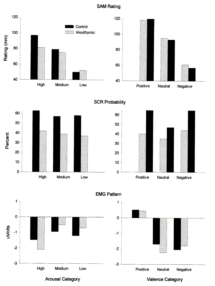
Self-Assessment Manikin. The twenty-one self-report ratings were placed into three groups of seven slides each for both affective dimensions based on the SAM ratings obtained from the control group. The means for both groups are presented in the top panels of Figure 1.
Both arousal and valence were characterized by linear, Fs(1, 61) = 118.61 and 471.54, respectively, ps < .001, and quadratic, Fs(1, 61) = 5.40 and 17.46, respectively, ps <.05 and < .001, trends across the three relevant categories. There was also a relationship between valence and arousal in this stimulus set (i.e. they were not completely orthogonal). Consistent with other studies (e.g. Fitzgibbons & Simons, 1992; Lang et al., 1993), slides with negative valence were most arousing, and neutral slides were least arousing, Flin(1, 61) = 471.54, p < .01, Fquad(1, 61) = 17.46, p < .01. There was no main effect for subject gender, though female subjects reported a wider range of positive and negative (i.e., valence) reactions to the slides, Gender X Valence Flin(1, 61) = 4.47, p < .05.
More importantly, alexithymic and control subjects differed in their self-reported emotion experience, and this difference was specific to the arousal dimension. A significant Group X Arousal interaction in the linear trend across categories supports the impression evident in Figure 1 that alexithymic subjects experience, or at least express verbally, a restricted range of arousal reactions, Flin(1, 61) = 6.59, p < .05. A test of the Group X Valence interaction did not approach statistical significance, Flin(1, 61) < 1.
Affective Words. Female subjects produced more affective words than male subjects, and, as predicted, alexithymic subjects produced fewer affect-related words than control subjects. Both differences were statistically significant, Gender F(1, 53) = 13.91, p < .01 and Group F(1, 53) = 4.03, p < .05, and held across all categories of emotion words. The difference between alexithymic and control subjects was due, in part, to a greater number of trials on which the alexithymic subjects failed to produce any words at all, F(1, 53) = 7.13, p < .01. The groups did not differ in their tendency to respond with slide, rather than emotion descriptors. Physiological ResponsesBoth heart rate and skin conductance responses were recorded over a one minute baseline. Controls and alexithymics had indistinguishable levels of tonic autonomic activity. The mean heart rate for control subjects was 75.00 beats per minute; for alexithymic subjects it was 74.50, F(1, 58) < 1. The mean number of non-specific skin conductance responses for control and alexithymic subjects was 2.47 and 1.73, respectively, F(1, 59) = 1.14, p > .25.
Skin Conductance Responses. The center panels of Figure 1 depict the relationships between SCR probability and the arousal and valence slide categories. As is typically the case (for review see Lang et al., 1993), there was a significant linear relationship between skin conductance and arousal, Flin(1, 58) = 5.22, p < .05, and a significant quadratic relationship between skin conductance and slide valence, Fquad(1, 58) = 20.12, p < .01. Also evident in the figures, alexithymic subjects were less responsive to the slides than were control subjects -- i.e alexithymic subjects produced fewer criterion SCR responses overall to the slide set, F(1, 58) = 4.98, p <.05. The patterns observed in the SCR magnitude data were similar to those in the probability data. Unlike the probability data, however, alexithymics and controls did not differ significantly in terms of overall magnitude, F(1, 58) = 1.01, p = .32. Thus, although alexithymics produced fewer responses, the responses they did produce were as large as those produced by control subjects.
Electromyographic Data. The relationship between the EMG pattern scores (Zygomatic - Corrugator) and both arousal and valence is displayed at the bottom of Figure 1. The facial pattern data were, as expected, directly related to stimulus valence, Flin(1, 53) = 8.65, p < .01, and Fquad(1, 53) = 7.70, p < .01, and not or only marginally related to arousal, Flin(1, 53) = 3.05, p = .09, and Fquad(1, 53) = 2.43, p = .12. As shown in the figures, the pattern scores of alexithymic and control subjects did not differ.
The linear relationship between valence and EMG data held for the corrugator, F(1, 56) = 9.95, p < .01, and zygomatic, F(1, 56) = 4.55, p < .05, muscles individually. The zygomatic-valence relationship was also quadratic, F(1, 56) = 7.71, p < .01, accounting for the quadratic observed in the pattern scores and most likely reflecting a 'grimace' in response to some of the negative slides (Bradley et al., 1993; Greenwald et al., 1989; Lang et al., 1993).
Heart Rate The mean heart rate response across all slides for alexithymic and control subjects is shown at the top of Figure 2. These waveforms are primarily deceleratory with an acceleratory component seen at mid interval. As the figure suggests, control subjects showed greater deceleration to the slides than did alexithymics, F(1, 57) = 8.50, p < .01. Males also showed greater overall deceleration to the slides than did females, F(1, 57) = 4.65, p < .05. There was no Group X Sex interaction. The subsequent acceleration in the waveform is the segment shown by Greenwald et al. (1989; see also Fitzgibbons & Simons, 1992) to be particularly sensitive to stimulus valence. As the bottom panels of Figure 2 illustrate, there was no significant relationship between HR acceleration and arousal (left), but positive slides were associated with a larger acceleratory component than were negative slides (right), Flin(1, 57) = 6.44, p < .05. The relationship between heart rate acceleration and valence was equivalent in the two subject groups, Flin(1, 57) < 1.
![]()
The basic self-report and psychophysiological data from this experiment are consistent with previous studies employing the same or similar methods (e.g., Bradley et al., 1992; Bradley et al., 1993; Greenwald et al., 1989; Lang, Bradley, & Cuthbert, 1990; Lang et al., 1993; Simons et al., 1993). The self-report data were similar to those of other studies incorporating the SAM, as was the relationship here between arousal and valence. Additionally, when the emotion-eliciting color slides are segregated into valence and arousal categories, the facial electromyogram data and the acceleratory component of the heart rate response were sensitive to stimulus valence whereas the skin conductance measures were related to arousal. The consistency of the data produced in this paradigm is impressive and gives added credence to the between-group findings. Likewise, confidence in the between-group effects is enhanced by the free-response self-report differences. By definition, alexithymic subjects have difficulty putting words to their emotion experience. In the present context, alexithymics provided fewer emotional descriptors than controls and for a greater number of slides produced no response at all.
More specifically, and as predicted, alexithymics appeared to experience both self-report and physiological deficits involving the arousal dimension of emotion and not involving the valence dimension. As mentioned above, skin conductance in the present investigation was a strong index of subject arousal. Alexithymic subjects in this experiment showed relatively fewer criterion skin conductance responses overall to the slide stimuli than did controls, and this finding paralleled the finding that alexithymics gave muted self-reports of arousal. The similarity between alexithymics= and controls= self-reports on the valence dimension replicates a finding from one of the only other studies which has asked subjects to rate their own feelings in terms of valence when they observed a group of stimuli varying in affective content (Wehmer et al., 1995).
On the other hand, the electromyographic data presented here are not consistent with the results reported by McDonald and Prkachin (1990). They found that during negative stimuli, alexithymics= facial expressions of emotion were less accurately and less intensely portrayed. This was not the case in the present study. One obvious source of variance between the present findings and those of McDonald and Prkachin lies in the specific measures of facial expression employed. Their judges rated the entire face of the subjects to determine the accuracy and intensity of emotions expressed. The present study looked only at activity in the corrugator (frown) and zygomatic (smile) muscles. Perhaps a wider sample of facial muscles would have been more reflective of the expressions studied by McDonald and Prkachin. The discrepancies between the two studies may also be due, in part, to the use of different instruments to identify alexythmia in their particpant pool. McDonald and Prkachin used the Shalling-Sifneos Personality Scale (Apfel & Sifneos, 1979) as opposed to the Toronto Alexythymia Scale that was employed in the present study.
Like facial EMG, heart rate acceleration in response to the slide stimuli was similar in the two groups. Because heart rate acceleration data and the electromyogram pattern scores are indices of valence, the fact that these measures did not differentiate alexithymics from controls is consistent with the fact that the groups= self-reports on the valence dimension did not differ. Indeed, the self-report and physiological responses of the alexithymics were concordant along both dimensions of emotion, suggesting that alexithymics= self-reports accurately reflected their affect-related responses.
Although alexithymics and controls did not differ in terms of heart rate acceleration, they did differ in terms of relative heart rate deceleration. This difference, however, was not valence related. Control subjects' heart rates decelerated more in response to slides of all categories. This would seem to indicate that alexithymics were less engaged by the slide stimuli. This interpretation is predicated on evidence which indicates that heart rate deceleration is characteristic of orienting or attention to input (Graham, 1979) and is consistent with an arousal-related deficit hypothesis, as arousal is generally highly correlated with self-reports of attention or interest (Lang et al., 1993). In fact, a recent study examining the effects of moving versus still pictures on affect-related physiological responses reached a similar conclusion (Detenber, Simons, & Bennett, 1997). In that study, picture motion did not change the relationship between heart rate acceleration and valence; picture motion was, however, associated with higher arousal ratings, larger SCRs, and greater heart rate deceleration than still pictures. This data pattern was interpreted by the authors as indicative of greater (sustained) attention allocated to the more arousing moving images.
If, as our data suggest, alexithymics are characterized by restricted arousal, this characteristic was not evident in the tonic measures obtained during the initial baseline. During baseline, alexithymics did not differ from controls in terms of absolute heart rate or number of spontaneous skin conductance responses. These findings are inconsistent with those of Wehmer et al. (1995) who found that alexithymics= heart rates were higher than controls= and interpreted this as supportive of elevated tonic sympathetic activity. Wehmer et al. recorded but did not report baseline skin conductance data. If, in fact, there were no between-group differences on their skin conductance measure, the data would not support the conclusion they drew. First, because heart rate is under both sympathetic and parasympathetic control, a higher basal heart rate need not reflect sympathetic influence. Second, subjects in Wehmer et al. were engaged in a task (creating stories to pictures) just prior to the baseline recordings which may well have had physiological consequences. Thus, their baseline was unconventional, rendering its interpretation difficult and a baseline comparison between experiments impossible.
arify the distinction between alexithymics and persons exhibiting a repressive coping style. Some authors have questioned this distinction (e.g., Martin and Pihl, 1986) based on evidence that both groups exhibit a dissociation ("decoupling"; Papciak et al., 1985) between physiological indices and reports of emotions. Repressors exhibit low self-reports of stress along with high stress-like physiological profiles in response to a stressor (Weinberger, Schwartz & Davidson, 1979). Likewise, alexithymics exhibit restricted affective self-report and experience--according to an emotional-discharge perspective--chronic hyperarousal. The present study's results, however, suggest that this similarity between groups may be more apparent than real; alexithymics appear to give accurate, rather than decoupled, accounts of their physiological state.
An assumption investigated in the present experiment was the possibility that although alexithymia is characterized by a deficit in the ability to describe feelings, the impact of this deficit may reach beyond vocabulary. The evidence presented in this paper sheds light on some of these effects. Alexithymics evince affect-related response characteristics which differentiate them from controls. Specifically, they exhibit physiological deficits which ostensibly substantiate their reports of experiencing less emotion than others and which indicate that this restriction most heavily involves the arousal dimension of emotions.
These data are particularly interesting when contrasted with other studies from our laboratory using similar procedures to study anhedonic subjects. In these studies (Fiorito & Simons, 1994; Fitzgibbons & Simons, 1992), anhedonic and control subjects differed only on valence-sensitive measures (SAM valence, heart rate and facial EMG), but no differences were noted on arousal self-report or skin conductance measures. Taken together, this set of studies demonstrates the utility of both the present paradigm and the dimensional conceptualization for the study of emotion. It also suggests that further studies involving these subject groups may shed light on the organization of emotion in general, and that such studies might include electroencephalographic measures as an attempt to define more clearly the nature of the arousal and valence deficits that characterize these interesting subject groups (e.g., Heller & Nitschke, 1997).
![]()
Abelson, R. P. & Sermat, V. (1962). Multidimensional scaling of facial expressions. Journal of Experimental Psychology, 63, 546-554.
Apfel, R. J. & Sifneos, P. E. (1979). Alexithymia: Concept and measurement. Psychotherapy and Psychosomatics, 32, 180-190.
Bagby, R. M., Taylor, G. J. & Atkinson, L. (1988). Alexithymia: A comparative study of three self-report measures. Psychosomatic Research, 32, 107-116.
Bagby, R. M., Taylor, G. J. & Parker, J. D. A. (1988). Construct validity of the Toronto Alexithymia Scale. Psychotherapy and Psychosomatics, 50, 29-34.
Berenbaum, H. & Prince, J. D. (1994). Alexithymia and the interpretation of emtion-relevant information. Cognition andEmotion, 8, 231-244.
Bradley, M. M., Greenwald, M. K., Petry, M. C., & Lang, P. J. (1992). Remembering pictures: Pleasure and arousal in memory. Journal of Experimental Psychology, 18, 379-390.
Bradley, M. M., Lang, P. J., & Cuthbert, B. N. (1993). Emotion, novelty, and the startle reflex: Habituation in humans. Behavioral Neuroscience, 107, 970-980.
Cacioppo, J. T., Uchino, B. N., Crites, S. L., Snydersmith, M. A., & Lang, P. J. (1992). Relationship between facial expressiveness and sympathetic activation in emotion: A critical review, with emphasis on modeling underlying mechanisms and individual differences. Journal of Personality and Social Psychology, 62, 110-128.
Cliff, N. & Young, F. W. (1968). On the relation between unidimensional judgments and multidimensional scaling. Organizational Behavior and Human Performance, 3, 269-285.
Detenber, B. H., Simons, R. F., & Bennett, G. G. (1997). Roll 'em!: The effects of picture motion on emotional responses. Journal of Broadcasting and Electronic Media, 21, 112-126.
Fiorito, E., and Simons, R. F. (1994). Emotional imagery and physical anhedonia. Psychophysiology. 31, 513-521.
Fitzgibbons, L. & Simons, R. F. (1992). Affective Response to color-slide stimuli in subjects with physical anhedonia: A three-systems anslysis. Psychophysiology, 29, 613-620.
Friedlander, L., Lumley, M. A., Farchione, T. & Doyal, G. (1997). Testing the alexithymia hypothesis: Physiological and subjective responses during relaxation and stress. The Journal of Nervous and Mental Disease, 185, 233-239.
Graham, F. K. (1979). Distinguishing among orienting, defense and startle reflexes. In H. D. Kimel, E. H. van Olst & J. F. Orlebeke (Eds.), The orienting reflex in humans (pp. 137-168). Hillsdale, NJ: Erlbaum.
Green, R. S. & Cliff, N. (1975). Multidimensional comparisons of structures of vocally and facially expressed emotion. Perception and Psychophysics, 17, 429-438.
Greenwald, M., Cook, E. & Lang, P. (1989). Affective judgement and psychophysiological response: Dimensional covariation in the evaluation of pictorial stimuli. Journal of Psychophysiology, 3, 51-64.
Heller, W. & Nitschke, J. B. (1997). Regional brain activity in emotion: A framework for understanding cognition in depression. Cognition and Emotion, 11, 637-661
Hyer, L. A., Woods, M. G., Summers, M. N. & Boudewyns, P. A. (1990). Alexithymia among Vietnam veterans with post-traumatic stress disorder. Journal of Clinical Psychiatry, 51, 243-247.
Jennings, J.R., Berg, W.K., Hutcheson, J.S., Obrist, P., Porges, S. & Turpin, G. (1981). Publication guidelines for heart rate studies in man. Psychophysiology, 18, 226-231.
Jessimer, M. & Markham, R. (1997). Alexithymia: A right hemisphere dysfunction specific to recognition of certain facial expressions? Brain and Cognition, 34, 246-258.
Kirkmayer, L. J. & Robbins, J. M. (1993). Cognitive and social correlates of the Toronto Alexithymia Scale. Psychosomatics, 34, 41-52.
Lane, R. D., Sechrest, L., Reidel, R., Weldon, V., Kaszniak, A. & Schwartz, G. E. (1996). Impaired verbal and nonverbal emotion recognition in alexithymia. Psychosomatic Medicine, 58, 203-210.
Lang, P. J. (1980). Behavioral treatment and bio-behavioral assessment: Computer applications. In J. B. Sidowski, J. H. Johnson, & E. A Williams (Eds.), Technology in Mental Health Care Delivery Systems (pp. 119-137). Norwood, NJ: Ablex.
Lang, P. J. (1985). The cognitive psychophysiology of emotion: Fear and anxiety. In A. H. Truma and J. D. Maser (Eds.), Anxiety and the anxiety disorders (pp. 131-170). Hillsdale, NJ: Erlbaum.
Lang, P. J., Bradley, M. M., & Cuthbert, B. N. (1990). Emotion, attention, and the startle reflex. Psychological Review, 97, 377-395.
Lang, P. J., Bradley, M. M., & Cuthbert, B. N. (1997). Motivated attention: Affect, activation and action. In P.J. Lang, R.F. Simons & M.T. Balaban (Eds.), Attention and orienting: Sensory and motivational processes (pp. 97-136). Hillsdale, NJ: Erlbaum.
Lang, P. J., Greenwald, M. K., Bradley, M. M. & Hamm, A. O. (1993). Looking at pictures: Affective, facial, and behavioral reactions. Psychophysiology, 30, 261-273.
Lang, P. J., Ohman, A. & Vaitl, D. (1988). The international affective picture system. [photographic slides]. Gainesville, FL: Center for Research in Psychophysiology, University of Florida, Gainesville.
Mann, L. S., Wise, T. N., Trinidad, A., & Kohanski, R. (1994). Alexithymia, affect recognition, and the five-factor model of personality in normal subjects. Psychological Reports, 74, 563-567.
Martin, J. B. & Pihl, R. (1985). The stress-alexithymia hypothesis: Theoretical and empirical considerations. Psychotherapy and Psychosomatics, 43, 169-176.
Martin, J. B. & Pihl, R. (1986). Influence of alexithymic characteristics on physiological and subjective stress response in normal individuals. Psychotherapy and Psychosomatics, 45, 66-77.
Martin, J. B., Pihl, R., Young, S. N., Ervin, F. R., & Tourjman, S. V. (1986). Prediction of alexithymic characteristics from physiological, personality, and subjective measures. Psychotherapy and Psychosomatics, 45, 133-140.
Mayer, J. D., DiPaolo, M. & Salovey, P. (1990). Perceiving affective content in ambiguous visual stimuli: A component of emotional intelligence. Psychosomatic Medicine, 54, 772-781.
McDonald, P. W. & Prkachin, K. M. (1990). The expression and perception of facial emotion in alexithymia: A pilot study. Psychosomatic Medicine, 52, 199-210.
Nemiah, J. C., Sifneos, P. E., & Apfel-Savitz, R. (1977). A comparison of the oxygen consumption of normal and alexithymic subjects in response to affect-provoking thoughts. Psychotherapy and Psychosomatics, 28, 167-171.
Newton, L. T. & Contrada, R. J. (1994). Alexithymia and repression: Contrasting emotion-focused coping styles. Psychosomatic Medicine, 56, 457-462.
Papciak, A. S., Feurestein, M. & Speigel, J. A. (1985). Stress reactivity in alexithymia: Decoupling of physiological and cognitive responses. Journal of Human Stress, 11, 135-142.
Parker, J. D. A., Taylor, B. J. & Bagby, R. M. (1993). Alexithymia and the recognition of facial expressions of emotion. Psychotherapy and Psychosomatics, 59, 197-202.
Rabavilas, A. D. (1987). Electrodermal activity in low and high alexithymia neurotic patients. Psychotherapy and Psychosomatics, 47, 101-104.
Reyes del Paso, G.A. & Vila, J. (in press). The continuing problem of incorrect heart rate estimation in psychophysiological studies: An off-line solution for cardiotachometer users. Biological Psychology.
Russell, J. (1980). A circumplex model of affect. Journal of Personality and Social Psychology, 39, 1161-1178.
Russell, J. & Mehrabian, A. (1977). Evidence for a three-factor theory of emotion. Journal of Research in Personality, 111, 179-183.
Schlosberg, H. (1952). The description of facial expression in terms of two dimensions. Journal of Experimental Psychology, 44, 229-237.
Sifneos, P. E. (1972). Short-term psychotherapy and emotional crisis. Cambridge, MA: Harvard University Press.
Simons, R. F., Fitzgibbons, L. & Fiorito, E. (1993). Emotion-processing in anhedonia. In N. Birbaumer & A. Ohman (Eds.), The Organization of Emotion (pp. 288-306). Toronto: Hogrefe-Huber.
Smith, C. A. & Ellsworth, P. C. (1985). Patterns of cognitive appraisal in emotion. Journal of Personality and Social Psychology, 48, 813-838.
Taylor, G. J. (1994). The alexithymia construct: Conceptualization, validation, and relationship with basic dimensions of personality. New Trends in Experimental and Clinical Psychiatry, 10, 61-74.
Taylor, G. J., Bagby, R. M. & Parker, J. D. A. (1997). Disorders of affect regulation: Alexithymia in medical and psychiatric illness. Cambridge: Cambridge University Press.
Taylor, G. J., Bagby, R. M., Ryan, D. P. & Parker, J. D. A. (1990). Validation of the alexithymia construct: A measurement-based approach. Canadian Journal of Psychiatry, 35, 290-297.
Taylor, G. J., Bagby, R. M., Ryan, D. P., Parker, J. D. A., Doody, K. & Deefe, P. (1988). Criterion validity of the Toronto alexithymia scale. Psychosomatic Medicine, 50, 500-509.
Taylor, G. J., Doody, K. & Newman, A. (1981). Alexithymic characteristics in patients with inflammatory bowel disease. Canadian Journal of Psychiatry, 26, 470-474.
Taylor, G. J., Ryan, D. & Bagby, R. M. (1985). Toward the development of a new self-report alexithymia scale. Psychotherapy and Psychosomatics, 44, 191-199.
Wehmer, F., Brejnak, C., Lumley, M. & Stettner, L. (1995). Alexithymia and physiological reactivity to emotion-provoking visual scenes. Journal of Nervous and Mental Disease, 183, 351-357.
Weinberger, D. A., Schwartz, G. E. & Davidson, R. J. (1979). Low-anxious, high-anxious, and repressive coping styles: Psychometric patterns and behavioral and physiological responses to stress. Journal of Abnormal Psychology, 88, 369-380.
![]()
1. Two studies have examined alexithymics' frontalis EMG activity in response to stress (Martin & Pihl, 1986; Papciak, Feuerstein & Spiegel, 1985). Although related, our focus is on EMG activity in response to emotion eliciting-stimuli, and our recordings are made over muscles of facial expression closely associated with the display of emotion.
2. Slides constituting the six discrete groupings are designated by their IAPS number as follows: Positive valence = 205, 451/421, 151, 720, 161, 501, 200; Neutral valence = 576, 160, 553, 691, 701, 166, 165; Negative valence = 104, 123, 221, 212, 451/421, 620, 300; Low arousal = 576, 161, 200, 501, 553, 701, 451/421; Medium arousal = 221, 151, 720, 104, 205, 123, 160; High arousal = 451/421, 691, 300, 212, 620, 165, 166. Note that slide 451 is a male nude and 421 is a female nude. Because slide ranking was determined across gender, the slide "451/421" in the positive valence and high arousal categories refers to slides depicting models whose sex is opposite that of the subject's and the slide 451/421 in the negative valence and low arousal categories refers to slides depicting models whose sex is the same as that of the subject.
4. All analyses were also performed with slide rankings determined by the average of alexithymic and control subjects' ratings. The findings were unchanged. .
![]()